News Story
Wonder "Worm"

Researchers developing mini robot for brain surgery
Removing any brain tumor is a delicate proposition. Deeply embedded tumors raise the stakes: Even the subtlest disturbance during neurosurgery can lead to disability or death.
An engineer, a radiologist and a neurosurgeon are combining their expertise to develop a tiny robotic tool that, aided by magnetic resonance imaging, one day could offer greatly improved visuals of brain tumors and pinpoint accuracy for removing them.
Jaydev P. Desai, University of Maryland associate professor of mechanical engineering who specializes in surgical robotics; Rao Gaullapalli, associate professor of diagnostic radiology and nuclear medicine; and neurosurgeon J. Marc Simard, both at the University of Maryland School of Medicine in Baltimore, head a research team that recently won $2 million from the National Institutes of Health to develop their prototype.
They’ve been working on the metal worm-like tool for several years in the kind of joint UMD-UMB research that the two institutions are encouraging through MPowering the State initiative.
"It allows our engineers and others to see firsthand the problems that physicians face, further motivating them to innovate new solutions in healthcare and design the next generation of biomedical devices," says Patrick O’Shea, UMD’s vice president for research and chief research officer.
Type, location and size of tumors are all factors in patients’ long-term outlook, as are advances in surgery, chemotherapy, and radiation treatments. The National Cancer Institute reported nearly 23,000 new cases of brain tumors in the U.S. in 2011, and 13,700 deaths.
In conventional neurosurgery, surgeons make a corridor that may be nearly as large as the tumor itself through normal overlying tissues. "Obviously, this is potentially harmful to the patient," says Simard. "It would be much better if a very narrow corridor could be made to introduce a miniature device to remove the tumor."
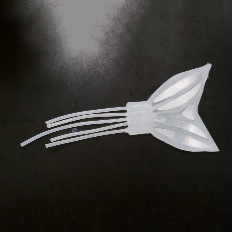
The Minimally Invasive Neurosurgical Intracranial Robot (MINIR) may be that device. About a half-inch in diameter and the length of a finger, MINIR would be inserted into the brain through a small opening. By watching continuous magnetic resonance images, the surgeon would be able to see the precise sites of the tumor and the robot.
The robot then electrocauterizes the tumor and sucks the debris through another suction tube, causing minimal damage to normal brain tissue.
"This technology has the potential to revolutionize the treatment of patients with difficult-to-reach tumors," Desai says. -- ET
(Reprinted with permission from TERP Magazine, Winter 2013, p. 38)
Desai is a member of the Maryland NanoCenter, affiliated with the Fischell Department of Bioengineering, and is director of the Robotics, Automation, and Medical Systems (RAMS) Laboratory.
Published May 1, 2013


